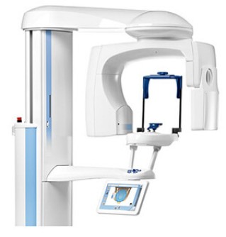
Digital volume tomography (DVT) uses X-rays to produce a three-dimensional image of your teeth.
In the same way as computed tomography (CT) or magnetic resonance imaging (MRI), DVT generates cross-sectional images of the area under examination. But in the DVT process – unlike CT – these cross-sectional images are produced by a three-dimensional X-ray beam and a two-dimensional image receptor. Digital processing then generates a 3D view from these cross-sectional images.
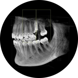
DVT makes it possible to visualise very fine structures inside the teeth that cannot always be clearly identified from conventional two-dimensional X-ray images.
With DVT, for example, it is possible to see the precise position of the wisdom teeth in relation to adjacent structures and also identify fractures and inflammations. In addition, DVT can be used to assess bone thickness and density, the direction of an implant, the proximity of the teeth to anatomical structures, and also restorative and aesthetic aspects.
This enables dentists to see whether bone augmentation is required. They can also use DVT for virtual planning of implant placements. For this reason, the method is sometimes known as "computer-guided implant surgery" (CGIS).
Another benefit: DVT helps dentists to plan minimally invasive interventions with considerably greater accuracy. They can assess the risks of treatment more precisely, provide more accurate prognoses and identify or exclude the causes of unclear disorders more reliably.
To summarise: DVT helps the dentist to achieve better results and avoid unnecessary interventions – including the associated costs.
DVT is only used for particular indications. In dentistry, these are:
Visualising complex root canal situations
Determining the positional relationships of resorptions, dental anomalies and supernumerary teeth
Planning dental implants
Visualising the positional relationship of wisdom teeth to nearby structures that they could endanger
Identifying inflammatory foci and bone pathologies
Investigating post-traumatic dental and bone injuries
Investigating the mandibular (jaw) joint
Visualising the paranasal sinuses and the adjacent structures